




Symptoms
- OSDI-6 score ≥ 4
The OSDI-6 keeps six of the original OSDI questions and is the screening questionnaire TFOS DEWS III recommends. Dry eye is always symptomatic, so a positive screen comes first.
ToolsOSDI-6 questionnaire
Learn · Dry eye diagnostics
A practical workup that follows the TFOS DEWS III Diagnostic Methodology (2025). Screen for symptoms, confirm lost homeostasis with a single objective sign, then pin down the drivers that shape treatment. Each step points to the test, the instrument and the supplies.
Making the diagnosis
TFOS DEWS III calls for a brief symptom questionnaire followed by at least one objective sign that tear film or ocular surface homeostasis has been lost.
The OSDI-6 keeps six of the original OSDI questions and is the screening questionnaire TFOS DEWS III recommends. Dry eye is always symptomatic, so a positive screen comes first.
The non-invasive method is preferred because it does not disturb the tear film. With fluorescein, the breakup time cut-off drops to < 5 s. If no osmometer is on hand, an unstable tear film is enough on its own.
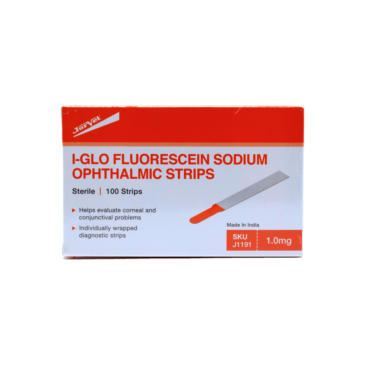
Use fluorescein for the cornea and lissamine green for the conjunctiva and lid margin. Both dyes can go on together.
Four stations in this order, so no test disturbs the tear film before the next one runs.
In the waiting room, before the exam starts.
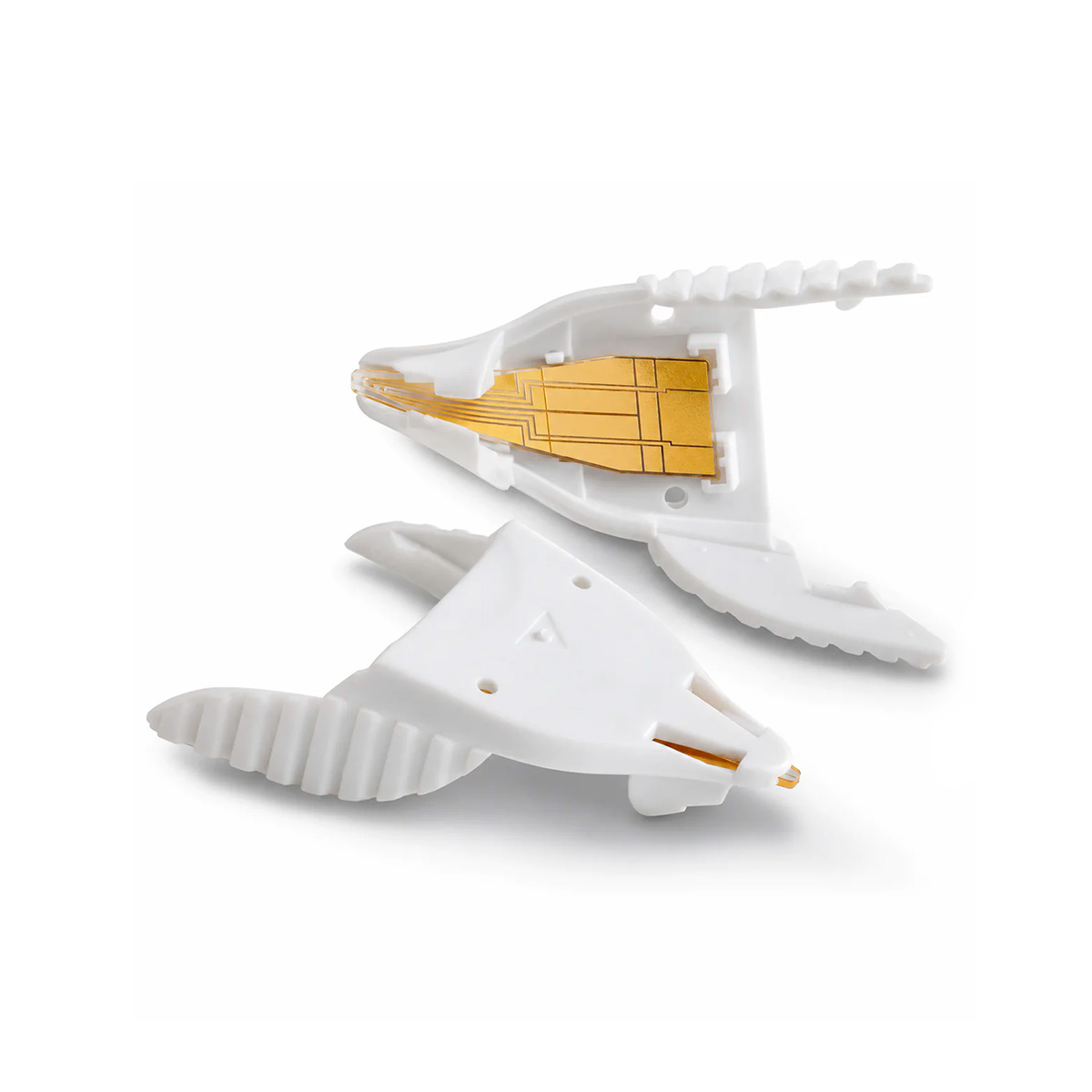
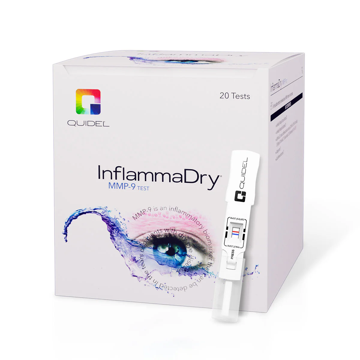
Collect tears before the slit lamp, any drops or any dyes.
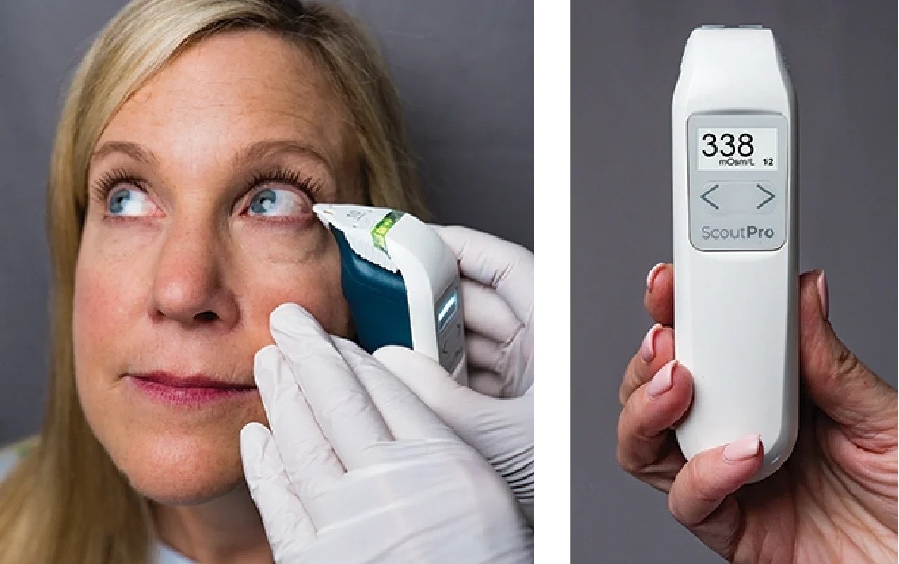
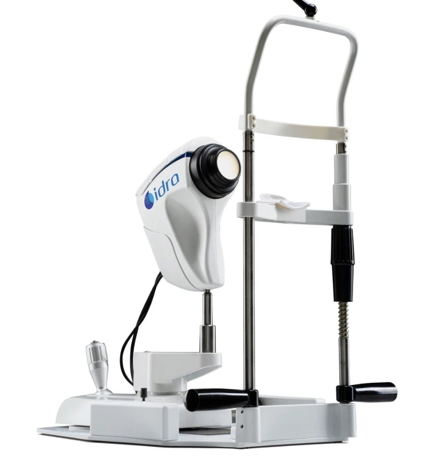
Non-contact and dye-free, on a single platform.
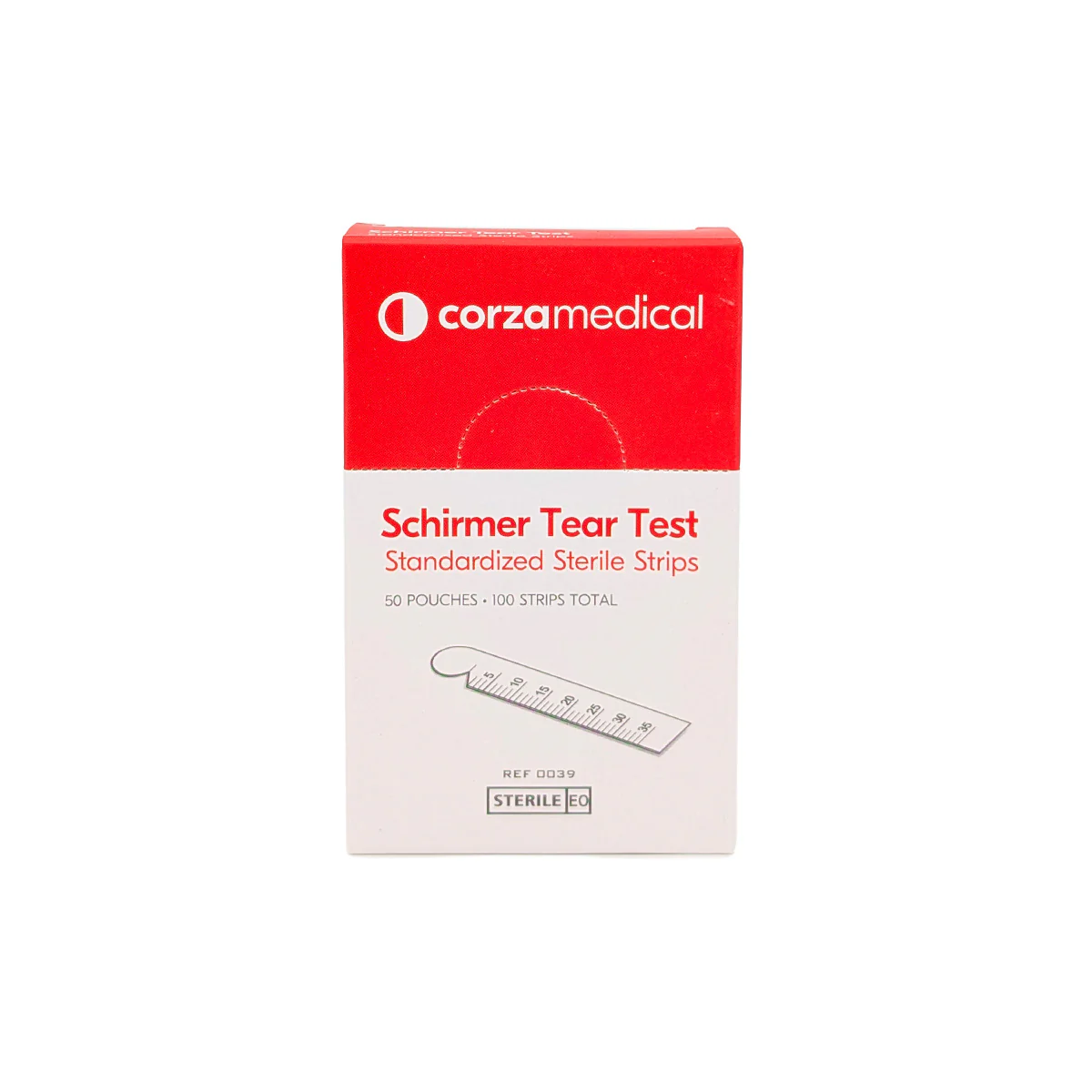
Contact and dye tests come last.
Order based on the TFOS DEWS III Diagnostic Methodology report.
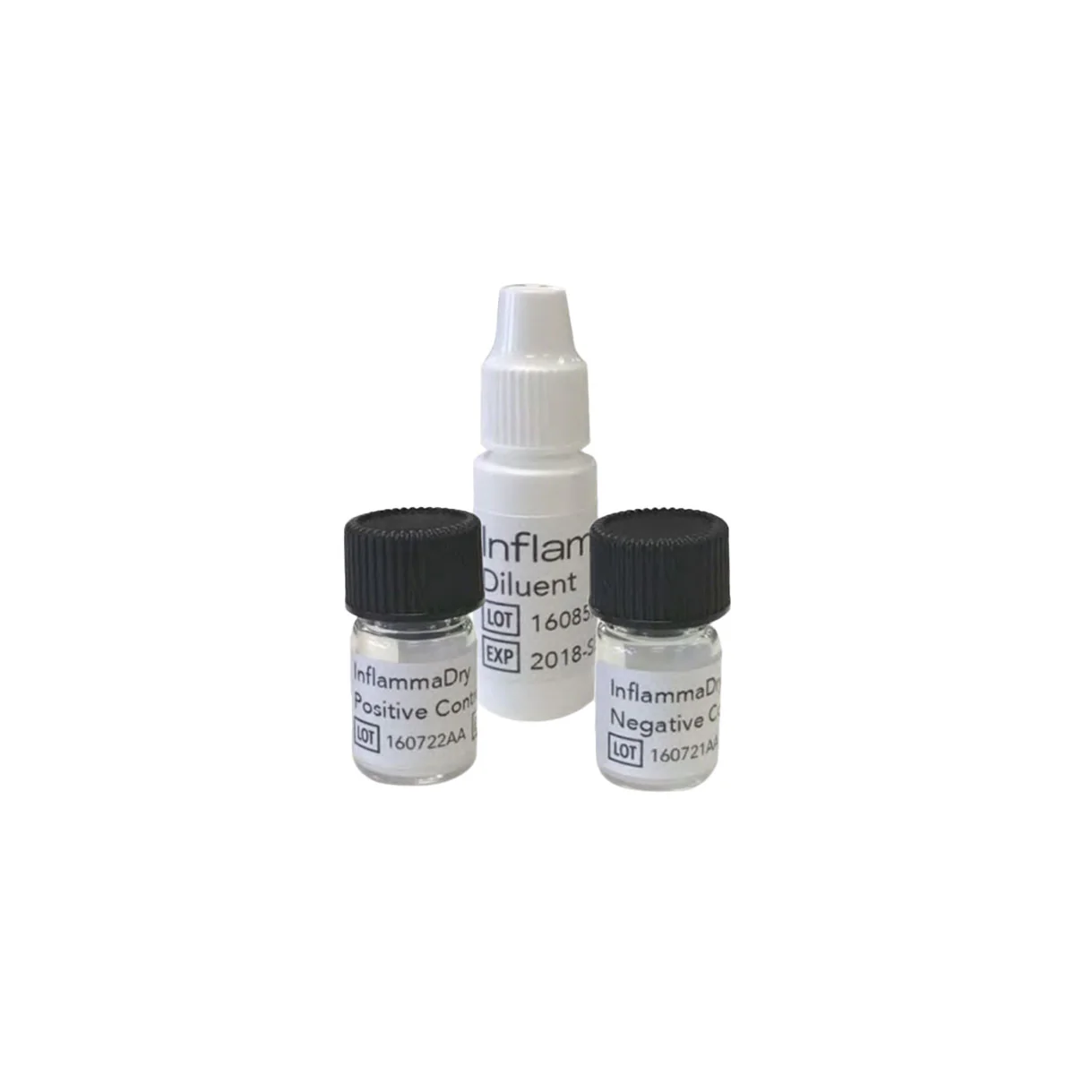
Both ScoutPro and InflammaDry need a tear film nobody has touched. Taking the osmolarity sample first and the MMP-9 sample second, straight after the questionnaire and before the slit lamp, satisfies both sets of instructions.
Subclassify
Rather than splitting dry eye into aqueous-deficient and evaporative, TFOS DEWS III classifies it by etiological driver. Most patients have several, and the dominant ones steer management.
Meibomian gland dysfunction and abnormal blinking thin the lipid layer and increase evaporation.
Reduced lacrimal gland output lowers tear volume, often because of systemic autoimmune disease.
Loss of goblet cells and damage to the epithelial glycocalyx destabilize the tear film at the surface.
Incomplete or infrequent blinks, a poor lid seal, and lid margin or eyelash disease.
Lid and globe misalignment, epithelial damage, primary inflammation and nerve dysfunction.
Autoimmune and endocrine disease, including Sjögren's disease, rheumatoid arthritis and lupus, as well as medications.
The point-of-care tests, dyes and strips this workup relies on, all from your Vision Rescue account.
Dry eye is now defined as a symptomatic disease involving lost homeostasis of the tear film, the ocular surface or both. The OSDI-6 with a cut-off of 4 becomes the standard screen, and subclassification looks for each patient's etiological drivers instead of an aqueous-deficient versus evaporative split.
A shortened Ocular Surface Disease Index made of the six questions that best separate dry eye from normal. It tracks the full OSDI closely and proved repeatable. A score of 4 or higher counts as a positive screen, and the full OSDI and DEQ-5 remain useful.
No. A positive OSDI-6 with a non-invasive breakup time below 10 seconds, or the staining criteria, is sufficient. Osmolarity is still valuable because hyperosmolarity sits at the core of the disease, and it gives you a number to track over time.
Sample before anything touches the tear film. For ScoutPro, allow 2 hours after drops or topical medication and 15 minutes after a slit lamp exam, anesthetic, dilating drops, invasive testing or crying, and never sample after staining. InflammaDry goes ahead of anesthetic, dyes or Schirmer strips. Osmolarity first and MMP-9 second, straight after the questionnaire, works for both.
InflammaDry detects elevated MMP-9 in tears and aids the diagnosis of dry eye alongside other clinical findings, adding information about inflammation. Topical or oral steroids, systemic immunomodulators, cyclosporine, tetracycline and topical azithromycin all suppress metalloproteinase activity and may lead to a false negative.
Thyroid eye disease, exposure keratopathy, allergic conjunctivitis, surface infection, ocular GVHD, contact lens complications, conjunctivochalasis, basement membrane dystrophy and neurotrophic keratopathy can all mimic dry eye. Because they can also occur alongside it, begin with a careful history and exam.
Age and sex, dry or drafty environments, screen and device use, autoimmune and endocrine disease, topical and systemic medications, contact lens wear, previous eye surgery, smoking and diet.
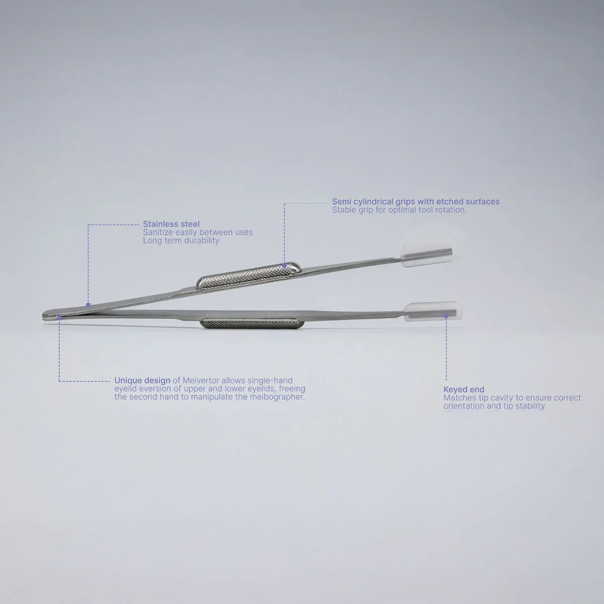
ScoutPro and its test cards, InflammaDry and its control set, fluorescein, lissamine green and Schirmer strips, the Meibomian Gland Evaluator and the Meivertor lid everter are on your wholesale account. IDRA, LipiScan and LipiView II and the Brill esthesiometer come directly from their manufacturers, and a lab processes the Sjö panel.
Begin with osmolarity, MMP-9 and the vital dyes, then add imaging as your dry eye patient base expands.
An educational summary of published diagnostic guidance for eye care professionals. It does not replace clinical judgment. Product names are trademarks of their respective manufacturers. Follow each manufacturer's instructions for use, including all warnings, precautions and contraindications.