









See gland health
View and assess the meibomian glands to support diagnosis and treatment decisions.
In-office procedures · Meibomian imaging
From Johnson & Johnson Vision, the LipiScan Dynamic Meibomian Imager and the LipiView II Ocular Surface Interferometer are office-based ophthalmic imaging devices for adults. LipiScan takes digital images of the meibomian glands, and LipiView II adds interferometric tear film imaging to measure the lipid layer.
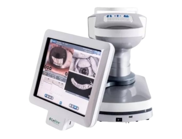
Each instrument images the meibomian glands using dynamic meibomian imaging. LipiView II goes further, recording specular (interferometric) observations of the tear film and measuring the absolute thickness of its lipid layer.
View and assess the meibomian glands to support diagnosis and treatment decisions.
LipiView II gives an absolute measurement of lipid layer thickness.
Images of the glands and tear film let patients see what is going on at the ocular surface.
LipiScan has a versatile design built to keep the exam lane moving.
LipiScan is dedicated to high-definition meibomian imaging, while LipiView II adds tear film interferometry and reporting to meibomian imaging.
A versatile unit for high-definition meibomian imaging, designed to keep the workflow efficient.
Patented interferometry that measures absolute tear film lipid layer thickness, along with imaging of the meibomian glands, eyelids and ocular surface.
Dual-Mode DMI, Dynamic Illumination and Adaptive Transillumination, three modes in all.
Real-time visualization through TearScience LipiView II Reporting.
Either device is run by a physician, who can capture images and then archive, edit and store them.
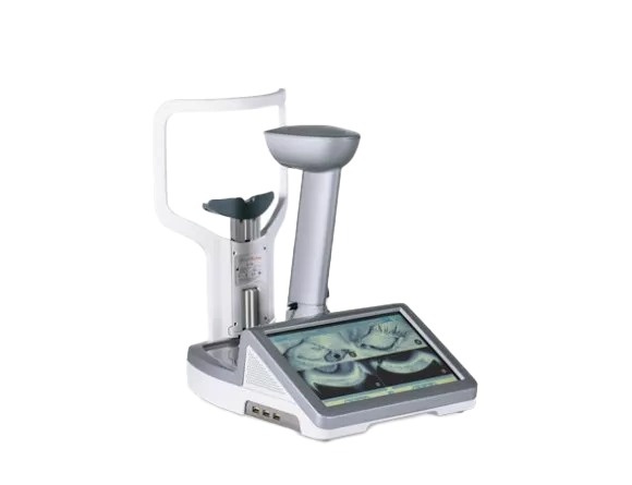
Dynamic meibomian imaging records the glands, and LipiView II adds the eyelids, ocular surface and tear film.
LipiView II uses those interferometric images to calculate how thick the lipid layer is, in absolute terms.
Saved images, plus real-time visualization in LipiView II Reporting, make review with the patient easy.
| LipiScan: device type | Ophthalmic imaging device |
|---|---|
| LipiScan: images | Digital images of the meibomian glands |
| LipiScan: imaging modes | Dynamic Illumination, Adaptive Transillumination, Dual-Mode DMI |
| LipiView II: device type | Ocular surface interferometer |
| LipiView II: tear film | Specular (interferometric) imaging; absolute lipid layer thickness |
| LipiView II: meibomian glands | Near-infrared (NIR) illumination |
| LipiView II: ocular surface and eyelids | White illumination |
| Patient population | Adults |
US: LipiScan: LipiScan™ Dynamic Meibomian Imager (DMI) is an ophthalmic imaging device intended for use by a physician in adult patients to capture, archive, manipulate and store digital images of the meibomian glands.
US: LipiView II: The LipiView II Ocular Surface Interferometer is an ophthalmic imaging device intended for use by a physician in adult patients to capture, archive, manipulate and store digital images of: • Specular (interferometric) observations of the tear film. Using these images, LipiView® II measures the absolute thickness of the tear film lipid layer. • Meibomian glands under near-infrared (NIR) illumination • The ocular surface and eyelids under white illumination
The meibomian glands, in digital images that can be saved, edited and archived.
Along with meibomian gland imaging, it uses interferometry of the tear film to measure lipid layer thickness in absolute terms, and it images the eyelids and ocular surface under white light.
Three in total: Adaptive Transillumination, Dual-Mode DMI and Dynamic Illumination.
A physician operates both, and both are intended for adult patients.
Use LipiScan to image the meibomian glands, or choose LipiView II to add lipid layer measurement of the tear film.
LipiScan, LipiView and TearScience are trademarks of Johnson & Johnson Vision. Read the complete manufacturer instructions for use, including every warning, precaution and contraindication, before use. Individual results vary.