









Classify the dry eye
Looks at each tear film layer and the glands together for a full assessment that helps pinpoint the type of dry eye.
In-office procedures · Dry eye diagnostics
IDRA analyzes each patient's tear film quickly and in structural detail. It looks at every layer, including lipid layer thickness and the meibomian glands, and is designed to help classify the type of dry eye and guide a treatment plan built for that patient.
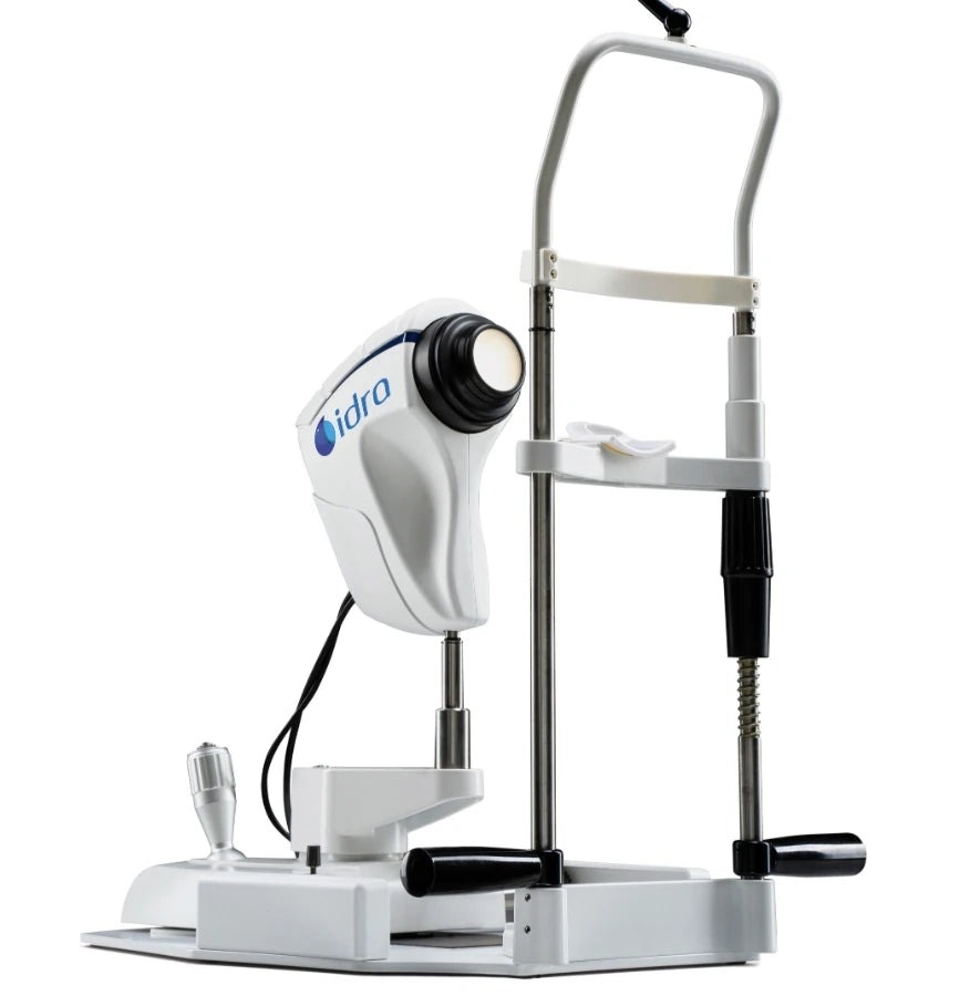
Automatic capture and analysis cover the lipid, aqueous and mucin layers plus the meibomian glands. The findings help classify the dry eye, and trained staff can run the assessment.
Looks at each tear film layer and the glands together for a full assessment that helps pinpoint the type of dry eye.
An automatic acquisition can take as little as 15 seconds.
The assessment is straightforward to hand off to trained staff.
Patients get documented findings and treatment options, and the 3D gland images make the conversation easier.
An algorithm proposes a possible approach for each patient, drawing on a list the doctor builds of the drugs, supplements and treatments the practice offers.
IDRA runs on the Instrument Control Platform (ICP), the software that captures and analyzes the surface exams.
Measures lipid layer thickness as a window on gland function, and can help in follow-up by picking up an increase in secretion after gland treatment.
One video yields an automatic NIBUT averaged across several readings, a stability-over-time graph and a topographic map of where the film breaks up.
Measures meniscus height automatically, a non-invasive indicator tied to aqueous production.
Three-dimensional gland images show lid structure and help confirm abnormal glands against a healthy reference.
An IDRA workup is quick, painless and non-invasive, and trained staff can perform it.
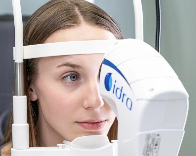
Infrared and visible light imaging record the tear film and ocular surface without contact.
Automatic captures can take as little as 15 seconds, and interferometry reports gland function within a few seconds.
Patient and data software puts images up for review right away, and the algorithm proposes a possible approach for each patient.
| Image resolution | 5 MP |
|---|---|
| Acquisition mode | Auto, manual; multi shot, video |
| Light sources | Infrared LED; blue, red and white LED |
| Sensor | Sensitive to infrared (NIR) |
| Cones | Main cone, Placido cone |
| Exams | Interferometry, tear meniscus, NIBUT, 3D meibography, blepharitis and cylindrical dandruff, bulbar redness, white-to-white, pupil diameter, anterior segment imaging, blink quality |
| Mini PC CPU | Intel Core i7-12700, 12 core, 2.10/4.90 GHz |
| Mini PC RAM | 16 GB DDR4 (3200 MHz) |
| Mini PC GPU | Intel UHD 770 4K, 2× DisplayPort, 1× HDMI |
| Connectivity | 2.5 Gb LAN, Wi-Fi 6, Bluetooth 5.2 |
| USB | 4× USB 3.2, 2× USB Type-C, 2× USB 2.0 |
| Operating system | Windows 11 Pro |
| Accessories | Smart Base, ophthalmic table |
Interferometry for lipid layer thickness, tear meniscus, NIBUT, 3D meibography, blepharitis and cylindrical dandruff screening, bulbar redness, white-to-white, pupil diameter, anterior segment imaging and blink quality.
An automatic acquisition can be done in as little as 15 seconds.
Yes. The assessment is built to be handed off.
Yes. Its algorithm proposes a possible approach for each patient from a list the doctor builds of the drugs, supplements and treatments the practice offers.
Reichert Technologies, through an exclusive US partnership with SBM Sistemi.
Tear film, meibomian gland and surface analysis, automated and combined on one platform.
IDRA is a trademark of SBM Sistemi, distributed in the US by Reichert Technologies. Read the complete manufacturer instructions for use, including every warning, precaution and contraindication, before use. Individual results vary.