









NIKBUT in seconds
Break-up time is measured automatically without dye, under infrared light that avoids glare.
In-office procedures · Dry eye diagnostics
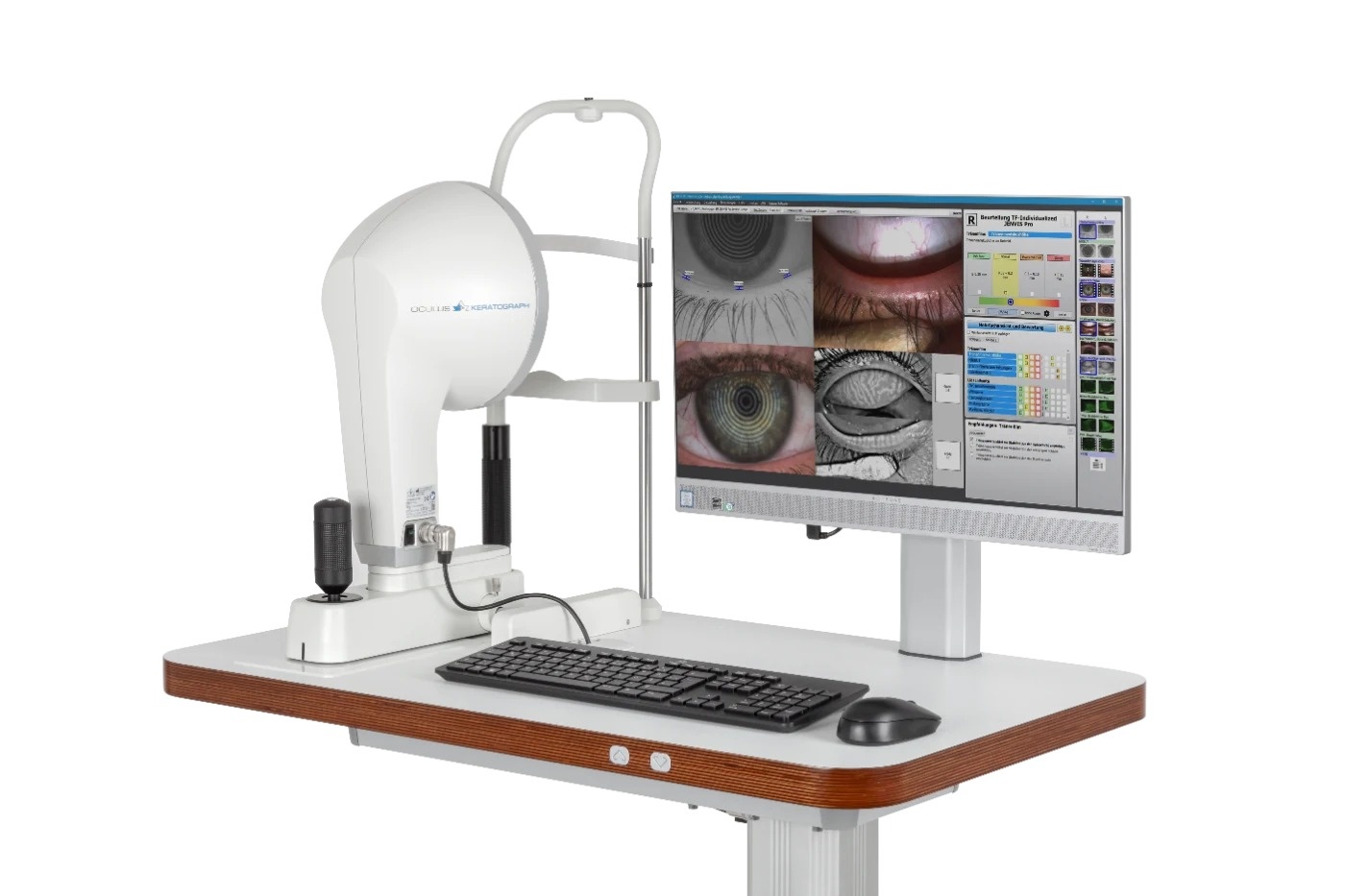
The OCULUS Keratograph 5M pairs an advanced corneal topographer and a true built-in keratometer with a color camera tuned for external imaging. In a dry eye workup it measures non-invasive tear break-up time, tear meniscus height, the lipid layer, the meibomian glands and bulbar redness.
Why practices add it
Using infrared and white light, the Keratograph 5M records tear film, lid and surface findings with no fluorescein. Calibrated workflows let technicians take the measurements, and automated analysis rolls everything into one report used for diagnosis, patient education and follow-up.
Break-up time is measured automatically without dye, under infrared light that avoids glare.
Meibo-Scan captures the upper and lower lid glands, and Meibo Analytics assesses gland structure and quantifies dropout automatically.
Tear meniscus height is measured without contact, and the lipid layer is judged from its interference pattern.
R-Scan grades bulbar and limbal redness on an objective scale.
The Crystal TEAR Report brings images, video and the patient's symptom answers into a single summary for the chart, patient education and treatment follow-up.
Placido topography, a true keratometer and several light wavelengths cover dry eye workups and contact lens fitting alike.
Twenty-two Placido rings produce 22,000 measured points across the anterior cornea, analyzed with Fourier and Zernike methods.
Reports measured anterior K values instead of simulated Ks.
White and 880 nm infrared Placido light, 465 nm blue light for fluorescein imaging and 840 nm infrared for meibography.
TF-Scan tear film analysis, Meibo Analytics, R-Scan, OxiMap oxygen transmissibility maps, pupillometry and a contact lens fitting database.
In the chair
The Keratograph 5M dry eye workup is non-invasive, so trained staff can perform it.
NIKBUT, meniscus height, lipid layer, meibography and redness are all taken with no dye and nothing touching the eye.
A four-test screening mode fits routine patients, NIKBUT takes only seconds, and tests run from least to most invasive.
Findings flow into the Crystal TEAR Report, and side-by-side views make follow-up visits and treatment tracking easier.
| Accuracy | ±0.1 D |
|---|---|
| Reproducibility | ±0.1 D |
| Placido rings | 22 |
| Measuring points | 22,000 |
| Working distance | 78–100 mm |
| Camera | Digital CCD camera |
| Light sources | Placido: white diodes and infrared diodes (880 nm); imaging: blue diodes (465 nm); meibography: infrared (840 nm); tear film dynamics: white diodes; pupillometry: infrared (880 nm) |
| Dimensions (W × D × H) | 275 × 320–400 × 480–510 mm |
| Weight, measuring head | 3.2 kg |
| Weight, with xy base | 6.1 kg |
| Max. power consumption | 18 W |
| Voltage / frequency | 100–240 V AC, 50/60 Hz |
| Data export | NIKBUT, R-Scan and keratometric data as CSV |
No. Non-invasive keratograph break-up time and fluorescein break-up time are measured differently and do not match exactly.
It is harder to measure when the corneal epithelium is badly damaged, and readings in those eyes tend to be very short.
Generally not, since the software detects distortion, but advanced keratoconus or marked irregular astigmatism can make the measurement difficult.
Meibo Analytics measures gland structure and dropout automatically, and the JENVIS Grading Scales let you compare images directly against reference grades.
It performs examinations that support a dry eye diagnosis.
No. Placido topography covers the anterior corneal surface only.
Yes. NIKBUT, R-Scan and keratometry data export to CSV.
One Keratograph 5M gives you corneal topography plus dye-free tear film, meibomian gland and redness testing.
Keratograph 5M is a trademark of OCULUS. Read the complete manufacturer instructions for use, including every warning, precaution and contraindication, before use. Individual results vary.